


+1-480-493-0093
Application Note 085
pdf (5.3 Mb)
Application Note 084
pdf (8.4 Mb)
Application Note 083
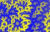
Atomic force microscopy (AFM) is routinely applied for compositional mapping of heterogeneous polymer materials. Recognition of the individual components in these materials is usually based on their specific morphology and differences of local mechanical and electric properties.
pdf (11.1 Mb)
Application Note 082
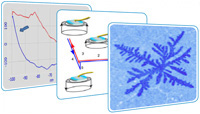
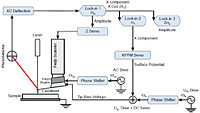
Sensitive measurements of local electrical properties, with a few nanometers spatial resolution, were realized in practice through phase modulation detection of the electrostatic force gradient. The validity of this approach is demonstrated on several different sample types: self-assemblies of fluoroalkanes, polymers, metals, and semiconductors.
pdf (2.8 Mb)
Application Note 081
pdf (2 Mb)
Application Note 080
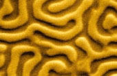
This short review describes the possibilities of imaging of biological materials using atomic force microscopy (AFM) in liquid environment. Atomic force microscopy can give new insight on biological matter, because it can work in environment close to native for the living cells, bacteria and viruses.
pdf (890 Kb)
Application Note 079
Demonstration of the pseudoatomic resolution in AFM imaging on open air is performed conveniently with a testing HOPG sample. The measurement procedure needs to use a sufficiently stiff and short cantilever and to define the load so that the pseudoatomic resolution was achieved both with the LAT signal distribution and with the DFL signal distribution, with the sample remaining non-destructed.
pdf (810 Kb)
Application Note 078
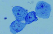
The investigation of biological objects, in particular, cells by scanning probe microscopy (SPM) demands a special preparing of biological object. This preparing means the fixation of the biological object.
pdf (360 Kb)
Application Note 077
As well known the classical resolution limit of conventional optical microscopes which arise from diffraction on entrancetive objective is equal to λ/2. It is deduced from approximation of flat waves, i.e. objective situating at the wave region of object radiation and light waves which come from this object may be considered as flat waves.
pdf (470 Kb)
Application Note 076
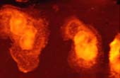
The work demonstrates on the biotin-streptavidin case abilities of SPM to be used for investigations of single molecule interactions. With using this technique some specific cases of binding were found.
pdf (380 Kb)
Application Note 075
Magnetic force microscopy (MFM) is effective tool to magnetic investigations on submicron scale. Image obtained by MFM is the space distribution of some parameter characterizing magnetic probe-sample interaction, i.e. interaction force, amplitude of vibrating magnetic probe etc.
pdf (220 Kb)
Application Note 074
During scanning in contact mode the cantilever bends not only along normally to the surface but also the cantilever torsional (lateral) deformation occurs. LFM measures the torsional deformation of the cantilever during scanning in contact mode.
pdf (360 Kb)

Application Note 073
The term “nanolithography” is commonly used for the local change of any properties of a surface by a scanning probe microscope (SPM) tip. It is the complex technique of creating and visualizing nanometer functional elements, including individual molecules and atoms, on a surface.
pdf (600 Kb)
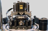
Application Note 072
Standard NT-MDT SI SPMs allow investigating a surface in temperature conditions from room one up to 150°C. The modified SMENA with heating stage provides the possibility of measurements with temperature up to 300°C.
pdf (300 Kb)

Application Note 071
Vacuum allows raising the Q-factor of cantilever oscillations, which, in its turn, substantially raises the sensitivity o light magnetic forces measurements between the probe and the sample.
pdf (570 Kb)
Application Note 070
Measuring in the magnetic field allows observing magnetic reversal processes and other effects that depend on the magnetic field. NT-MDT SI devices allow carrying out measuring in the longitudinal and perpendicular magnetic fields. The strength of the magnetic field is controlled by the build-in Hall effect sensor.
pdf (500 Kb)
Application Note 069
Measurements carried out with heating or cooling of the sample allows studying the changes of sample’s properties with the variations of temperature. There are several capabilities of carrying out the investigations with the varying temperatures in the air and in liquids in NT-MDT SI equipment.
pdf (600 Kb)