NTEGRA II – Modular SPM (AFM, STM) system | NT-MDT S.I.
+1-480-493-0093

NTEGRA II


Leading AFM in Nanoscale Analysis

Brochure

  • Leading AFM in Nanoscale Analysis
  • Atomic Force Microscopy with all the latest solutions
  • Jumping AFM – HybriD ModeTM
  • Automation of the experiment – ScanTTM
  • Widest possibilities for specific cases
  • Open architecture system
  • User-friendly software

Key features

NTEGRA II – Precise, Fast and Intelligent Microscope

NT-MDT II introduces NTEGRA II, a second generation of the most popular AFM in the world.

With added capabilities, enhanced functionalities, it delivers an unprecedent level of modularity and flexibility becoming a true partner of a researcher.

Intelligent, fast, reliable, precise and, undeniably, easy to use.

To phase out influence of noisy environments, NTEGRA II comes with a standard enclosure that provides temperature control, acoustic and vibration isolation.

Such combination reveals a true nature of NTEGRA II as the most stable AFM in the world that delivers a thermal drift at less than 0.2 nm/min level.


Intelligent ScanT™

Program has been developed with the help of neural networks to provide auto-tuning of scanning parameters for imaging in AM-AFM.

Automatic maintenance of attractive (non-contact) & repulsive (intermittent-contact) regimes.

Artefact-free scanning without parachuting. Perfect performance at samples with any kind of morphology.

To learn more


Al2O3 with ScanTTM


Fast RapidScan™

NTEGRA II enables scans for up to 10 Hz with standard cantilevers (resonance frequency <500 kHz) and up to 25 Hz with short cantilevers (resonance frequency >1 MHz).

No additional hardware and software are required.

To learn more


90×90×0.5 μm image of collagen fibers. 1&10Hz scanning rates


Open design & easy access to the sample

Open architecture in hardware, software, and signal integration provides a portal for you to customize and expand this list to fit your applications.

Until nowadays NTEGRA concept remains unsurpassed. It was designed specifically to form an AFM based platform within cutting edge methods from other scientific analyses such as raman spectroscopy and nanoIR spectroscopy.

NTEGRA II create a core around which your lab can build a full spectrum of analytical operations.

NTEGRA II offers an elegant solution. Universal platform design, universal electronics, universal software.

No matter what the configuration, NTEGRA II will retain its excellent quality and performance.


Base unit NTEGRA II


HybriD Mode™

The HybriDTM is now an AFM standard mode that enables quantitative nano-mechanical measurements to provide researches with Young modulus maps, maps of adhesion, deformation, etc.

Furthermore, implementation of non-oscillatory technique in electrical modes, such as conductive AFM, PFM, KPFM, allows now researchers to work with fragile and loosely fixed samples.

In HybriD mode (US Patient 9,110,092) the tip-sample distance is modulated according to the harmonic law. Thus tip enters a force interaction with the sample thousands times per second.

High-performance electronic components and unique algorithms implemented in the state-of-the-art HybriD 2.0 Control Electronics provide superb level of real-time signal processing and analysis. Combining mode with cutting-edge optical microscopy and spectroscopy techniques opens-up novel opportunities of cantilever-based tip-enhanced Raman scattering (TERS) and scattering scanning near-field optical microscopy (s-SNOM).

To learn more


HybriD ModeTM – scanning technique based on fast force-distance curves measurements with real-time processing of tip response


NT-MDT NOVA PX – Software suit

Spectra Analysis

Spectrum processing in real-time & offline.

Wavelength change in a single click.

Lasers, gratings, polarizers, pinholes and so on, are fully automated & controlled from the single software.

Integration with spectra databases


Image Analysis

More than 100 methods for AFM-Raman data processing & analysis

Powerful toolkit for AFM image leveling

Smart package for automatic practical detection Nonlimited number of copies under onsite license.

ISO-ASME-compatible statistical analysis.


Programmed configuring module

The module is a powerful tool for controlling the AFM measuring system.

The main element of the module is an interac- tive block diagram of the device.

With its help, the operations of organizing the work- ing block diagram of the device, generation, syn- chronous detection and switching of signals, control of digital filtering, organization of feedback systems, etc. are carried out.

When working according to the standard methods included in the NOVA PX program, all switches in the controller are carried out automatically when the measurement method is selected.


AFM Heads


SMENATM

Scan AFM head for measurements in liquid and gas environment in basic AFM modes.

Scanner: Scanning range – X,Y,Z 100х100х10 um.

AFM Z noise down to 20 pm.

Cantilever oscillations registration - up to 5 MHz.

Bias voltage – 500 kHz.

Tip-to-sample manual approach option.

Registration system: 650 nm (optionally 1300 nm) or SLD 850 nm.

Exchangeable probe holder for operation in liquid.


SNA 3

AFM-Raman head optimized for top/side/bottom illumination.

Manual probe registration system.

AFM Z noise down to 15 pm.

Optical access from top/side for objectives (WD>6 mm) or condenser (NA<0.3).

Optical access from bottom for objectives (including immersion objectives).

Includes removable AFM unit for cantilever probe.

Registration system 670 nm (optionally 830 or 1300 nm).


Extensions

NTEGRA Spectra II – AFM-Raman

Change happens at interfaces and today’s most exciting changes in microscopy are happening where multiple technologies interface.

NTEGRA Spectra II is a prime example, uniting the full power of confocal microscopy, atomic force microscopy, Raman & fluorescence spectroscopy in one platform.

From topography to spectrum analysis, from electrical and mechanical properties to optical spatial resolution below the diffraction limit.


AFM topography

TERS (D band)

AFM topography

TERS (D band)
TERS + HybriD ModeTM of Graphene Oxide on Au substrate. TERS resolution: 10 nm


Life Biology

NTEGRA II combines the strengths of the excellent AFM with perfect optical microscope for biological and medical applications.

From molecules to living organisms, NTEGRA II opens the window to the world of biology and biochemistry. Especially designed to integrate seamlessly with your optical microscope, NTEGRA II preserves the in situ environment so that you can observe, image and measure what is really there.

To maintain life and present the best conditions for measurement, most biological samples must be kept in fluid solutions.

For conventional AFM biological imaging as well as biochemistry and bioorganic applications, NTEGRA II uses a unique sealed fluid cell which maintains an enclosed volume.


Fragment of Musca domestica eye. Size: 50x50x7,3 um

Triangle-shaped DNA origami scanned in liquid. Scan size: 2,5x2,5 um

Amiloid-like fibrils of CSD protein in the process of their formation by Y-Box binding protein (YB-1) on mica surface. Scan size: 10x10 um

Seleno-protein deposited on mica from solution. You can see some organic parts and molecules of proteins on the image. Scan size: 5x5 um


Vacuum & External field NTEGRA II

NTEGRA II is a high-sensitive system perfectly suited for measurements in vacuum up to 10-3 torr or under controlled atmosphere environments.

The widespread application AFM with enhanced MFM measurement capabilities in external magnetic field.


HybriD ModeTM-KPFM of WS2 flakes grown on epitaxial graphene overlaid over topography. Scan size: 8x8 μm

MFM maps of Yttrium Iron Garnet film before (left) and after (rigth) application of in-plane magnetic field. Scan size: 30x30 μm

KPFM of WS2/graphene heterostructures in ambient. Potential range 0.58 V. Scan size: 5x5 μm

KPFM of WS2/graphene heterostructures in vacuum (1×10⁻⁶ mbar). Potential range 1.77 V. Scan size: 12x12 μm

Applications

  • Biology and Biotechnology
  • Materials Science
  • Semiconductors
  • Polymers and Thin Organic Films
  • Data storage devices and medias
  • Piezoelectrics & Ferroelectrics
  • Nanocomposites
  • Carbon materials
  • Nanowires & Nanotubes
  • Domain structures


AFM Modes

Contact AFM: LFM, FMM, SRI, PFM
AM-AFM: MFM, EFM, SCFM, KPFM
HybriD ModeTM: Young’s modulus, Work of Adhesion, Current, Force Volume, PFM, KPFM, MFM, EFM, Thermal Microscopy
AFM spectroscopy, SS PFM Nanolithography: Voltage, Current, Force Scanning Tunneling Microscopy


Controlled Environment

Vacuum
Temperature
Gases
Liquid
Electrochemical environment
External magnetic field Humidity


E. coli in liquid
AM-AFM

TGS crystal near Curie temperature
HybriD ModeTM-PFM

Thin film of semiconducting polymer on Si
AM-AFM

sPS particles on Si
ScanTTM


DNA Origami
ScanTTM

Garnet Film
MFM

F14H20 on HOPG
AM-AFM

PAH Synthetic Nanographene
AM-AFM

Specifications

Scan type Scanning by sample Scanning by probe
Sample size Up to ⌀40 mm, up to 15 mm in height Up to ⌀100 mm in diameter, up to 15 mm in height
Sample weight Up to 100 g Up to 300 g
XY sample positioning range, resolution 5x5 mm, 5 μm
Positioning sensitivity 2 μm
Scan range 100x100x10 μm 100x100x10 μm
Up to 200x200x20 μm (DualScanTM mode)
Non linearity, XY ≤ 0.1% ≤ 0.15%
Noise level, Z
(RMS in bandwidth 1000 Hz)
0.04 nm (typically), 0.06 nm 0.06 nm (typically), 0.07 nm
Noise level, XY
(RMS in bandwidth 200 Hz)
0.2 nm (typically), 0.3 nm (XY 100 um) 0.1 nm (typically), 0.2 nm (XY 50 um)
Optical viewing system Optical resolution 3 μm
(1μm optional;
0.4 μm optional, NA 0.7)
3 μm
Field of view 4.5-0.4 mm 2.0-0.4 mm
Continuous zoom available available
Vibration isolation Active  0.7-1000 Hz

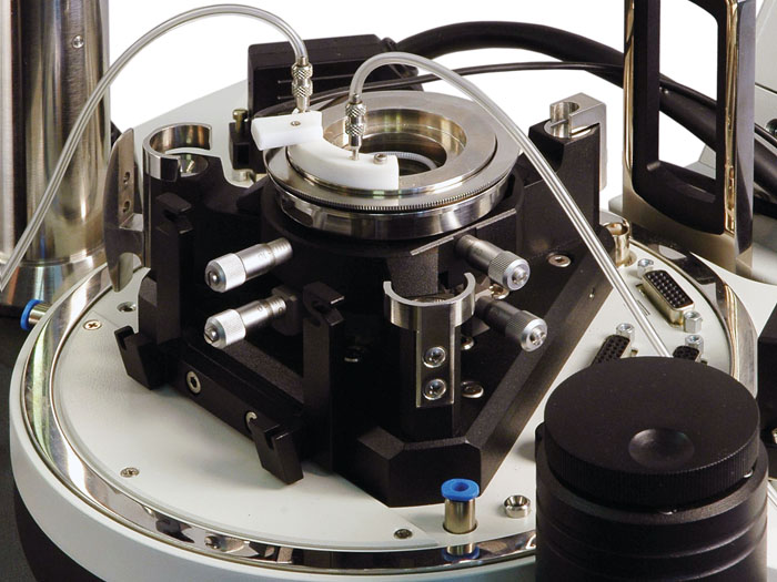
Measuring modes and techniques

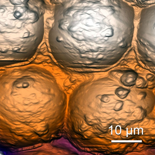
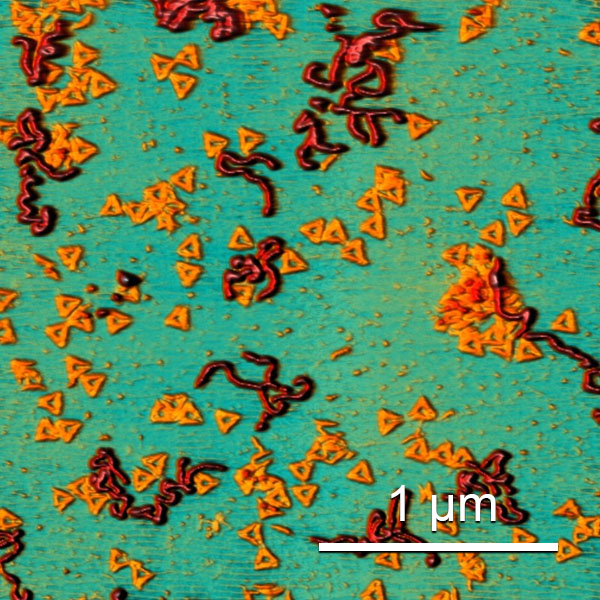
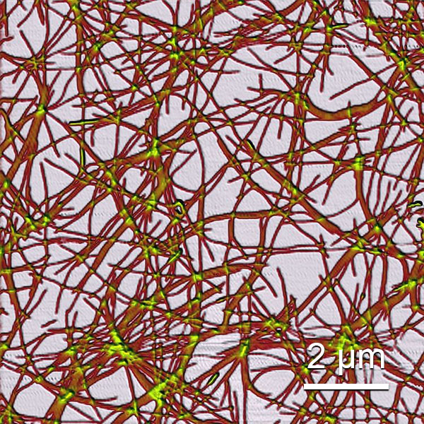
Contact AFM: Topography, Lateral Force, Force Modulation, Spreading Resistance Imaging
Amplitude modulation AFM: Topography, Phase, Feedback
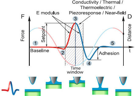
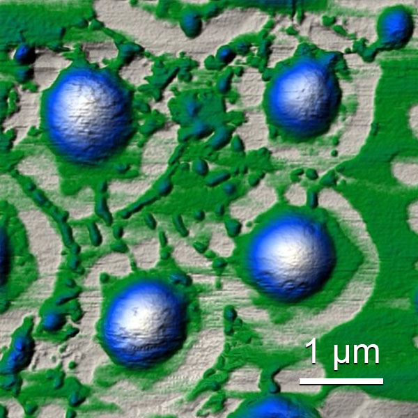
HybriD ModeTM AFM: Topography, Young’s modulus, Work of Adhesion, Viscoelectisity, Current, Piezoresponse Force Microscopy, Fast Force Volume
AFM spectroscopy: Force-distance, Amplitude-distance, Phase-distance, I(V), I(Z)
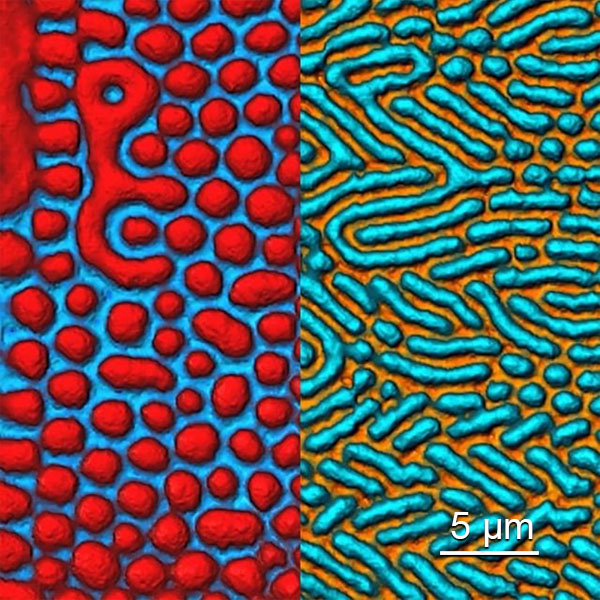
Magnetic Force Microscopy: Two-pass and Frame Lift DC/AC
Electrostatic Force Microscopy: Single-pass and Two-pass Amplitude Modulation, Frequency Modulation
Scanning Capacitance Force Microscopy: Single-pass and Two-pass Amplitude Modulation, Frequency Modulation (dC/dZ and dC/dV imaging)
Kelvin Probe Force Microscopy: Single-pass and Two-pass Amplitude Modulation, Phase Modulation
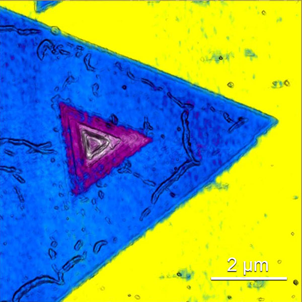
Piezoresponse force microscopy & Switching Spectroscopy
Nanolithography: Voltage, Current, Force

More information


Information brochures

Key publications

  • Kumari, P., Arora, N., Chatrath, A., Gangwar, R., Pruthi, V., Poluri, K. M., & Prasad, R. "Delineating the biofilm inhibition mechanisms of phenolic and aldehydic terpenes against cryptococcus neoformans". ACS Omega 2019, 4, 18, 17634–17648 https://doi.org/10.1021/acsomega.9b01482
  • Dubrovin, E. V., Barinov, N. A., Schäffer, T. E., & Klinov, D. V. "In Situ Single-Molecule AFM Investigation of Surface-Induced Fibrinogen Unfolding on Graphite". Langmuir 2019, 35(30), 9732–9739 https://doi.org/10.1021/acs.langmuir.9b01178
  • Herma, R., Wrobel, D., Liegertová, M., Müllerová, M., Strašák, T., Maly, M., Semerádtová, A., Štofik, M., Appelhans, D., & Maly, J. "Carbosilane dendrimers with phosphonium terminal groups are low toxic non-viral transfection vectors for siRNA cell delivery". International Journal of Pharmaceutics, 562 (2019), 51–65 https://doi.org/10.1016/j.ijpharm.2019.03.018
  • Yevdokimov, Y. M., Skuridin, S. G., Salyanov, V. I., Bobrov, Y. A., Bucharsky, V. A., & Kats, E. I. "New optical evidence of the cholesteric packing of DNA molecules in “re-entrant” phase". Chemical Physics Letters, 717, (2019), 59–68 https://doi.org/10.1016/j.cplett.2019.01.017
  • Kaysheva, A. L., Pleshakova, T. O., Stepanov, A. A., Ziborov, V. S., Saravanabhavan, S. S., Natesan, B., Archakov, A. I., & Ivanov, Y. D. "Immuno-MALDI MS dataset for improved detection of HCVcoreAg in sera". Data in Brief, 25, 2019, 104240 https://doi.org/10.1016/j.dib.2019.104240
  • Obraztsova, E. A., Basmanov, D. V., Barinov, N. A., & Klinov, D. V. "Carbon Nanospikes: Synthesis, characterization and application for high resolution AFM". Ultramicroscopy 197, 2019, 11–15 https://doi.org/10.1016/j.ultramic.2018.11.004
  • Koplak, O., Kravchuk, Useinov, Talantsev, A., Hehn, M., Vallobra, P., Mangin, S., & Morgunov, R. "Surface engineering of magnetic and mechanical properties of Ta/Pt/GdFeCo/IrMn/Pt heterostructures by femtosecond laser pulses". Applied Surface Science, 2019, 493, 470–477 https://doi.org/10.1016/j.apsusc.2019.07.024
  • Koplak, O. V., Sidorov, V. L., Kunitsyna, E. I., Valeev, R. A., Korolev, D. V., Piskorskii, V. P., & Morgunov, R. B. "Bistable and Multi-Domain States of α-Fe/(PrDy)(FeCo)B Ferromagnetic Microwires". Physics of the Solid State, 2019, 61, 2061–2068 https://doi.org/10.1134/S1063783419110209
  • Tavares, M. A. B., Andrade, L. H. F., Martins, M. D., Gomes, G. F. M., Fernandez-Outon, L. E., & Matinaga, F. M. (2019). "Precession damping in [Co60Fe40/Pt]5 multilayers with varying magnetic homogeneity investigated with femtosecond laser pulses". AIP Advances,2019, 9, 125322 https://doi.org/10.1063/1.5130458
  • Figueiredo-Prestes, N., Oliveira, R. C., Tavares, M. A. B., Costa, D. S., Mazzaro, I., Jurca, H. F., Zarpellon, J., Martins, M. D., Deranlot, C., George, J. M., & Mosca, D. H. "Stabilization and tuning of perpendicular magnetic anisotropy in room-temperature ferromagnetic transparent CeO2 films". Journal of Applied Physics, 2019, 126, 183903 https://doi.org/10.1063/1.5125321
  • Prokopov, A. R., Mikhailova, T. V., Danishevskaya, E. V., Shaposhnikov, A. N., Berzhansky, V. N., Karavainikov, A. V., Nedviga, A. S., Nauhatsky, I. A., & Milyukova, E. T. "Bi-Substituted Iron Garnet Films for Thermomagnetic Recording, Photonics, and Plasmonics: Optimization of Synthesis Conditions Using Scanning Probe Microscopy". Technical Physics, 2019, 64(11), 1709–1715 https://doi.org/10.1134/S1063784219110239
  • Chirkov, N. S., Akentiev, A. V., Campbell, R. A., Lin, S. Y., Timoshen, K. A., Vlasov, P. S., & Noskov, B. A. "Network Formation of DNA/Polyelectrolyte Fibrous Aggregates Adsorbed at the Water-Air Interface". Langmuir 2019, 35(43), 13967–13976 https://doi.org/10.1021/acs.langmuir.9b02487
  • Horvath, I., Blockhuys, S., Šulskis, D., Holgersson, S., Kumar, R., Burmann, B. M., & Wittung-Stafshede, P. "Interaction between Copper Chaperone Atox1 and Parkinson’s Disease Protein α-Synuclein Includes Metal-Binding Sites and Occurs in Living Cells". ACS Chem. Neurosci. 2019, 10, 11, 4659–4668 https://doi.org/10.1021/acschemneuro.9b00476
  • Sofińska, K., Wojtkiewicz, A. M., Wójcik, P., Zastawny, O., Guzik, M., Winiarska, A., Waligórski, P., Cieśla, M., Barbasz, J., & Szaleniec, M. "Investigation of quaternary structure of aggregating 3-ketosteroid dehydrogenase from Sterolibacterium denitrificans: In the pursuit of consensus of various biophysical techniques". Biochimica et Biophysica Acta - General Subjects, 1863 (2019), 1027–1039 https://doi.org/10.1016/j.bbagen.2019.03.009
  • Morozova, O. V., Levchenko, O. A., Cherpakova, Z. A., Prokhorov, V. V., Barinov, N. A., Obraztsova, E. A., Belova, A. M., Prusakov, K. A., Aldarov, K. G., Basmanov, D. V., Lavrenova, V. N., Pavlova, E. R., Bagrov, D. V., Lazarev, V. N., & Klinov, D. V. "Surface modification with polyallylamines for adhesion of biopolymers and cells". International Journal of Adhesion and Adhesives, 92 (2019), 125–132 https://doi.org/10.1016/j.ijadhadh.2019.03.013
  • Parshina, E. Y., Yusipovich, A. I., Brazhe, A. R., Silicheva, M. A., & Maksimov, G. V. "Heat damage of cytoskeleton in erythrocytes increases membrane roughness and cell rigidity". J Biol Phys 45, 367–377 (2019) https://doi.org/10.1007/s10867-019-09533-5

Contact Us

Fulfill a special form to request additional information.

Get more information