


+1-480-493-0093


Atomic Force Microscopy is a widely used and powerful technique for investigating materials at a nanoscale range. However, the technique is not simple to use, which elicits varied results between researchers with different levels of experience. NT-MDT Spectrum Instruments (formerly NT-MDT) have created the intelligent software, ScanT™, inspired by neural networks to make dynamic amplitude modulation AFM (AM-AFM) easy for researchers of every skill level.
The AFM amplitude modulation (AM) mode, also known as “tapping” or “semi-contact”, is based on dependency of cantilever amplitude oscillation on distance between sample surface and tip. This mode is often favored over other AFM modes because it provides minimal impact on the sample and the tip, which can help to preserve both.
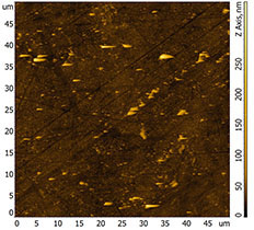
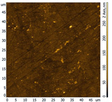
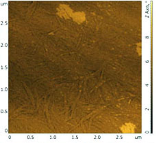
However, in practice a large portion of AFM images contain artefacts of different kinds, including uncontrollable switching of cantilever oscillation regime, extra noise of measured signals (ringing) or detachment the probe from the surface (parachuting effect) (fig.1). These distortions can affect the interpretation of the results following analysis and can in turn lead to incorrect conclusions based on those results.
To achieve a high image quality the user must manually and simultaneously manipulate major parameters during the scan to avoid artefacts including, set point, tip oscillation amplitude, scanning speed and feedback gain. This is especially important for rough surface, objects weekly connected to the surface (nanoparticles, nanotubes, single molecules), soft samples (gels, soft polymers, biological objects), or samples where the topography is unknown before the scan.
This not only requires a good understanding of the microscope, the principles of how it works and how different parameters will affect the image quality in different scenarios, but also experience with these different types of samples to reliably produce acceptable images. All this does not influence well to the efficiency of the AFM.
This presents another hurdle as gaining experience requires access to samples and the time needed to learn the equipment and the scanning technique.
As a response, automation of the parameter tuning needed for image acquisition in AM-AFM is highly desirable for users to avoid spending lots of time and efforts on mastering the instrument.
The intelligent ScanT™ software allows the user to get quickly high-quality and reliable results on their samples in AM ACM.
The ScanT™ application has been developed with the help of neural networks to provide auto-tuning of scanning parameters for imaging in AM-AFM. Requiring minimal knowledge about sample properties and minimal involvement from the end user, this software is useful not only for beginners but also to assist experienced researchers, especially where the topography of the sample is unknown beforehand.
The ScanT™ software reduces noise level to a minimum, significantly reduces image defects of the topography such as parachuting, feedback excitation, and switching between attraction and repulsion tip-surface interaction regimes, and provides high quality imaging of samples despite different roughness and material nature.
The ScanT™ algorithm identifies defects during the tuning procedure and eliminates or reduces them through feedback gain optimization, as well as adjusting the set point, tip oscillation amplitude and scanning speed.
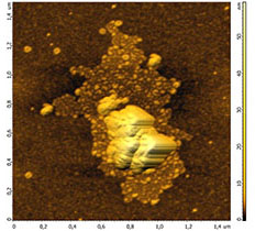
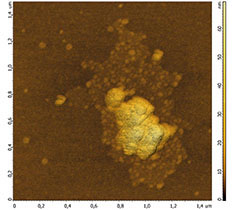
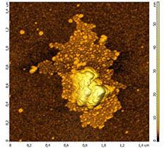
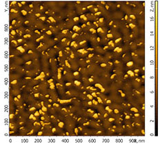
Figure 2 shows an example of non-optimal feedback setting (left and center) in comparison with the image, when feedback gains are optimally selected.
One the most common defects in AM-AFM are caused by choosing the wrong set point and probe oscillation amplitude that stimulate in turn switching the tip oscillation from an attractive to a repulsive regime.
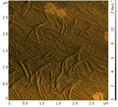
The topography of these images looks “torn” and in many cases together with low quality results can guide a researcher to the wrong conclusions. ScanT™ removes or significantly minimizes these types of defects with confident force control that automatically tunes the set point and amplitude to fit the sample and allows scanning only in attractive or repulsive regimes. Figure 3 shows examples of images obtained in the attraction regime and when it jumps between attraction and repulsion occur.
One of the advantages of scanning in an attractive regime is the preservation of tip sharpness, increasing the life time of probe considerably. However, presence of the topography with steep slopes on the surface, like large particles, runs the risk of crushing the probe during the scan. ScanT’s algorithm monitors the feedback error signal and doesn’t allow the tip to be crushed during tuning and scanning.
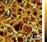
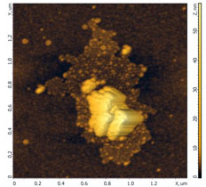
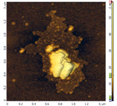
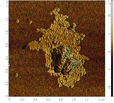
The ScanT™ software can also confidently control the low sample-tip interaction force in an attraction regime. ScanT™ makes the imaging of tricky and soft samples repeatable and routine (fig.4).

The module has a user-friendly interface which allows the straight forward launching of the imaging procedure (Fig.5). To run a measurement, an attractive or repulsive scanning regime is chosen and then the scan is started.
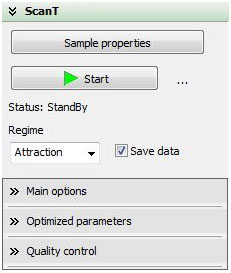
At the same time all image quality indicators and advanced controls are still available for experts who want to be kept informed during the scan.
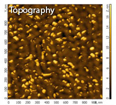
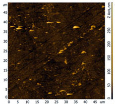
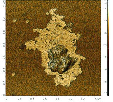
If the surface has features with more or less steep borders, defects on their images can occur due to tip detachment from the surface (parachuting effect). It happens fairly often when the image of almost entire scanned surface is of high quality and only a few small areas, usually associated with individual particles, have such defects. As a result, the quality of this image becomes unacceptable and re-scanning of this area is required.
The GTransformTM algorithm built into the program allows eliminating the “parachuting” defects in the topography image. As a consequence, an acceptable result can be obtained from the image of the moderate quality (see fig. 5).
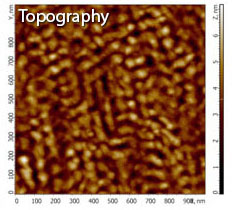
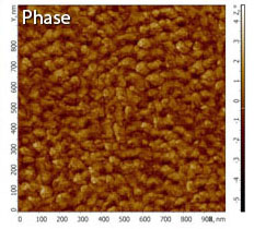
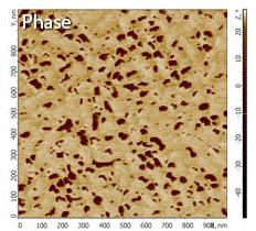
In addition, the GTransformTM algorithm allows extracting substantial information from a scanned image. Indeed, the images of phase contrast, spreading current or lateral forces contain, along with information related to the heterogeneity of the surface properties, also the contrast induced by the topography. In some cases this can significantly mask the map of measured surface property heterogeneity. Figure 6 shows how GTransformTM removes or significantly reduces the distortions of phase contrast images caused by surface topography making contrast produced by material property inhomogeneity more pronounced.
For beginners the smart ScanT™ software provides an easy gateway into producing high quality AFM images, with one-click optimization and without the steep learning curve to understand all the details and gain enough experience to create reliable/trustworthy images.
For experts the ScanT™ software provides assistance to free up time that would otherwise be spent producing rudimentary images, without sacrificing control over the procedure, and allows researchers to widen the range of materials that they routinely scan into tricky, delicate or soft materials, thus increasing the potential scope of their research.
pdf (9.2 Mb)