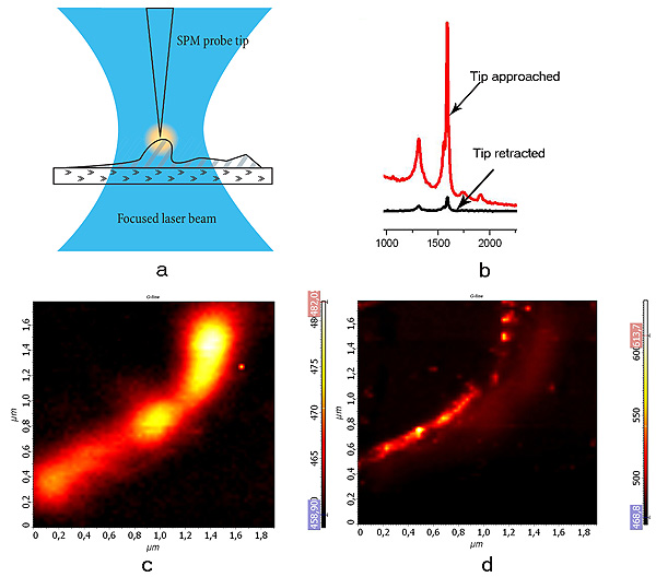
Raman microscopy with ultra-high spatial resolution. a - tip enhanced Raman scattering experiment, b - intensity of carbon nanotube G-band increases by several orders of magnitude when the probe tip is landed, c - confocal Raman image of carbon nanotube bundle. d- tip-enhanced Raman scattering (TERS) image of the same nanotube bundle. Note, TERS provides more than 4-times better spatial resolution as compared to confocal microscopy.
Data courtesy of Prof. Dr. G. de With, Dr. S.Kharintsev, Dr. G. Hoffmann, Dr. J. Loos, TUE, the Netherlands.