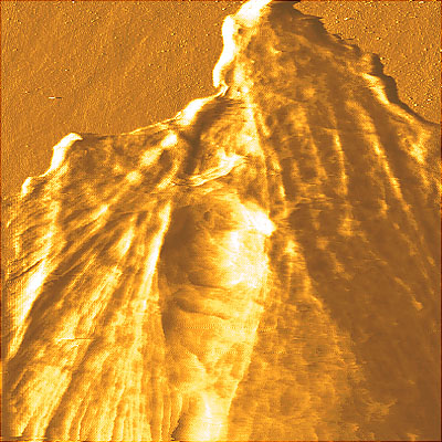
Contact error mode AFM image of a part of living porcine kidney proximal tubule epithelial cell (LLC-PK1). The cytoskeleton of the cell is clearly visible. Image was obtained in the contact mode in a buffer solution at 37C.
Sample courtesy of Prof. Tang Ming-Jer, Department of Physiology
National Cheng Kung University Medical College, Tainan, Taiwan (ROC).