NTEGRA Marlin
+1-480-493-0093

NTEGRA MARLIN


Cutting-edge AFM-RAMAN-SICM System for Biological Studies
Project is supported by The Foundation for Assistance to Small Innovative Enterprises (FASIE)

Cutting-edge AFM-RAMAN-SICM System for Biological Studies

Brochure

  • AFM-SICM-Raman: triple synergy of powerful nanocharacterization techniques
  • Hopping mode ion conductance microscopy:
    non-contact imaging of living cells and jelly surfaces
  • QNM: power of AFM and SICM quantitative nanomechanical studies
  • Smart Patch Clamp: automatic nanopipette targeting for ion channel studies
  • High-Speed Long-Range High-Resolution mapping at nanoscale
  • Nano-injection: nanopipettes with SICM feedback control can be used
    for local sub-picoliter injections to single cells

SICM Principle

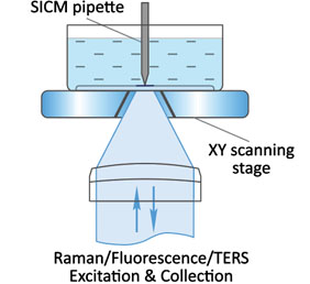
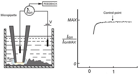
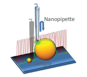
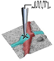
SICM (Scanning Ion Conductance Microscopy) is an SPM technique which uses nano-pipette (sharp glass electrode) for non-contact 3D surface mapping at high resolution. In SICM, the probe to sample distance is controlled via the decrease of ionic current flowing through the tip, as it approaches the sample surface.
Biophys.Journ. 73, 653-658


a)

b)

c)

d)
Continuous and “Hopping” SICM images of highly corrugated neuron cell

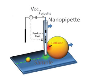
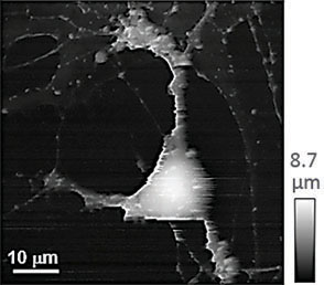
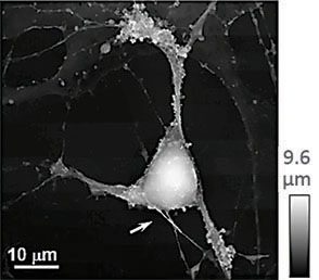
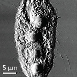
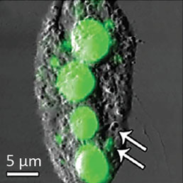
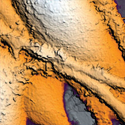
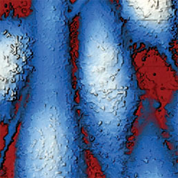
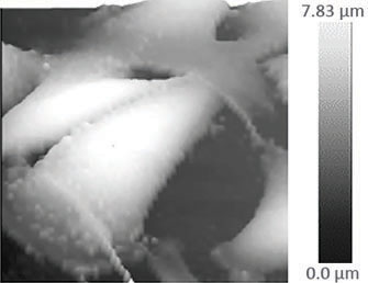
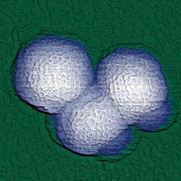
(a) Illustration of a scanning nanopipette probe operating in continuous scan mode colliding with a spherical object possessing a steep vertical slope. (b) Illustration of the hopping mode used in HPICM showing how the pipette is withdrawn to a position well above the sample before approaching the surface. (c,d) Topographical images of the same fixed hippocampal neuron obtained first with hopping mode (d) and then with continuous left-to-right raster scan mode (c), using the same nanopipette.
Hopping mode algorithm applied to SICM allows to image uneven and convoluted samples at high resolution ensuring that pipette always approaches from above rather than “dragging” along the surface. Nature Meth. (2009) 6: 279-281

Recording of the webinar presented by Prof. Yuri Korchev (Imperial College London): “NTEGRA Marlin: Bringing SICM to Your Research”.

SICM imaging of living cells

Noncontact algorithm of hopping SICM enables stable, fast and high-resolution imaging of soft and highly corrugated objects like living cells under physiologically relevant conditions.
Scanning method ensures that the probe is always approaching the sample in vertical direction, thus, it becomes possible to visualize even those objects that are “suspended” in space.


Neuron from mouse hippocamp 10×10×6,3 μm

B16 melanoma cells 25×25×5,4 μm

PC3 human prostatic carcinoma cells 40×40×6,8 μm

SICM image of live neuron from mouse hippocamp 40×40×13.3 μm

Smart patch clamp

Smart patch-clamp combines conventional patch-clamp and SICM.
SICM generates a high-resolution topography followed by ion current recording in a specified location.

Neuron (2013) 79. 1067-77. Nanoscale-Targeted Patch-Clamp Recordings of Functional Presynaptic Ion Channels

Extracellular pH mapping of single living cells

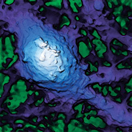
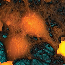
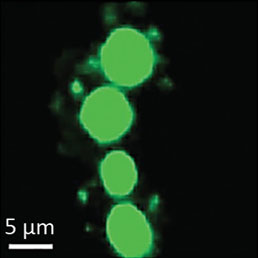
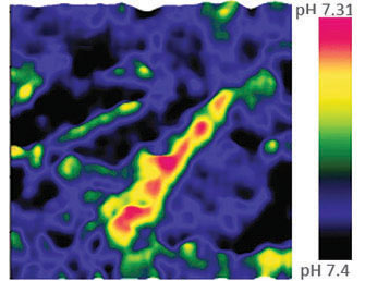
Extracellular pH mapping of living cancer cells with high spatial resolution and sensitivity can be implemented by means of SICM utilizing doublebarrel nanopipette. Morphology and pH-map of low-buffered living melanoma cells are shown from the left. Scale bars represent 20 μm.
Nature Comm. (2019) 10, 5610

High Resolution AFM

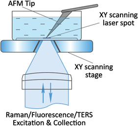
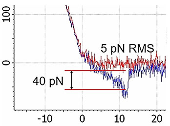
High-resolution AFM microscopy is available by means of Contact, Tapping and HybriD modes and is empowered by lowest signal-tonoise ratio of OBD loop on the market down to 25 fm/√Hz.


200×200×100 nm high-resolution AFM topography of rhinovirus particles

Quantitative Nanomechanical Studies (QNM)

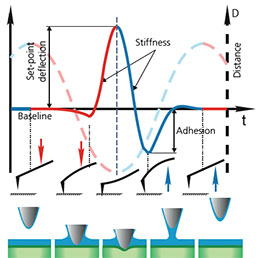
Combination of SICM and HybriD Mode™ AFM expands the boundaries of real-time quantitative nanomechanical mapping to 10 orders of elastic modulus keeping the possibility of single-point force spectroscopy experiments. Low- or noninvasive nature of tip-sample interaction allows to study delicate biological and jelly samples that are weakly attached to the substrate.


SICM E-Modulus map of live fibroblast cell. SICM E‑modulus map of live fibroblast 25×25 μm E=2 Pa..3,4 MPa

Streptavidin-biotin affinity single-molecule detection

HybriD AFM E‑modulus map of stem cell in HybriD Mode 18×18 μm E=200 kPa..50 MPa

HybriD Mode™

Correlative Imaging

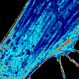
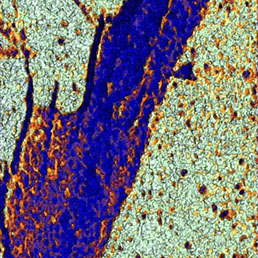
Flawless hardware and software integration of AFM/SICM with confocal Raman/fluorescence microscopy provides the widest range of additional information about the sample. Simultaneously measured AFM/SICM and Raman/fluorescence maps of exactly the same sample area deliver complementary information about sample physical properties and chemical composition.
When equipped with specially prepared probes, which are working as “nanoantennas”, AFM/SICM-Raman combination allows to perform optical mapping with resolution less than diffraction limit and is called TERS: Tip Enhanced Raman Scattering.


a)

b)

c)
Combined AFM-Raman microscopy studies of cyanobacterial film. AFM Phase map (a). Raman map showing the intensity distribution of the Raman band at 1521 cm-1 corresponding to beta-carotene (c). Raman-AFM overlay (b). Typical Raman spectrum of the sample containing a band at 520 cm-1 that is the Si-Si stretching mode of the silicon AFM tip and bands at 1160 cm-1 and 1521 cm-1 assignable to beta-carotene

Specifications

SICM

  • Position control: capacitive closed-loop sensors on X,Y,Z axes
  • XY travel range: 30×30 μm (optionaly up to 100×100 μm
  • Z travel range: 25 μm
  • Z position accuracy: 0.1 nm
  • Applications: SICM (Hopping mode), SECM, Smart Patch-Clamp, Microinjection, QNM, pH mapping
  • Typical image acquisition time: less than 5 minutes (depends on point number and sample roughness)

AFM

  • XYZ closed-loop tip scanner 100×100×10 μm
  • High-performance low noise AFM: Z noise <0.1 nm (RMS in 10-1000 Hz bandwidth)
  • Measurements in gas and liquid environment
  • Applications: all standard AFM imaging modes (40+), Force-distance spectroscopy
  • Non-resonant oscillatory HybriD Mode™ allowing direct and fast force detection for QNM mapping (E-modulus, Adhesion, Deformation, etc.)

Optical Spectroscopy

  • Confocal Raman/fluorescence/Rayleigh imaging runs simultaneously with AFM/SICM
  • Diffraction limited spatial resolution: <200 nm in XY, <500 nm in Z (with immersion objective)
  • True confocality; motorized confocal pinhole for optimal signal and confocality
  • Continuously variable ND filter with the range 1 - 0.001 for precise change of laser power
  • Motorized variable beam expander/collimator: adjusts diameter and collimation of the laser beam individually for each laser and each objective used
  • Fully automated switching between different lasers - with few mouse clicks
  • Full 3D (XYZ) confocal imaging with powerful image analysis
  • Extremely high efficiency 520 mm length spectrometer with 4 motorized gratings
  • Visible, UV and IR spectral ranges available
  • Echelle grating with ultrahigh dispersion; spectral resolution: 0.007 nm (<0.1 cm-1)
  • Up to 3 different detectors can be installed:
    - TE cooled (down to -100 °C) CCD/EMCCD cameras
    - APD in photon counting mode or FLIM detector
    - PMT for fast confocal laser (Rayleigh) imaging
  • Flexible motorized polarization optics in excitation and detection channels, crosspolarized Raman measurements
  • Low wavenumber/THz Raman spectroscopy: <10 cm-1 with Bragg volume filters
  • Hyperspectral imaging (recording complete Raman spectrum in every point of 1D, 2D or 3D confocal scan) with further software analysis
  • Highest possible resolution optics is used simultaneously with AFM/SICM: up to 1.45 NA
  • Dual scan: scan by sample AND scan by laser spot (for Hot Spot mapping in TERS)
  • Closed-loop scanning mirrors for precise laser spot positioning to the tip (important for SNOM, TERS)

Contact Us

Fulfill a special form to request additional information.

Get more information